Educational Workshop- Gastric Ultrasound
Miguel Magallanes Esqueda, MD
Liana Victoria Zucco, MD
Issue 3 | Volume 2 | August 2026
OVERVIEW
The department’s workshop series has drawn on faculty, fellows, and residents across several subspecialties.
Dr. Liana Victoria Zucco coordinated the major gastric ultrasound workshop, which had promising stations and consisted of a dynamic and interacive day.
May 22: Ultrasound workshop led by Michael Li.
June 24: Ultrasound workshop spearheaded by Michael Li.
July 8: Education Resources Workshop.
Additional contributors: Peter Santer, Matthew Gao, Matthew Heron, Sahil Amin
Simulation colleagues: Nadav Levy, Federico Puerta Martinez, and Yitzhak Brzezinski Sinai contributed on the day as well.
The Center for Education Research, Technology and Innovation (CERTAIN) within the Department of Anesthesia, Critical Care and Pain Medicine at Beth Israel Deaconess Medical Center hosted a multidisciplinary educational workshop on perioperative gastric ultrasound on July 22, 2026. The workshop was developed to strengthen learners’ understanding of gastric ultrasound as a point-of-care tool for aspiration risk assessment by combining foundational educational material with hands-on image acquisition, clinical decision-making, and immersive simulation.
PARTICIPANTS AND FORMAT
Participants included anesthesiology interns, residents, fellows, faculty anesthesiologists, and medical students. Before attending the workshop, learners completed introductory educational content through the department’s learning management system (Docebo), including interactive Articulate Storyline modules and foundational presentations covering gastric anatomy, image acquisition, and the clinical application of gastric ultrasound. During the workshop, participants rotated through five educational modules designed to reinforce both technical and clinical competencies. These modules focused on assessment of clear gastric contents, dynamic evaluation of gastric emptying, and clinical management of aspiration risk and GLP-1 receptor agonist therapy, together with immersive virtual reality simulation. The workshop concluded with three five-question knowledge assessments that allowed participants to reinforce key concepts and receive immediate feedback.
WORKSHOP MODULES
Module 1. Model With Empty Stomach
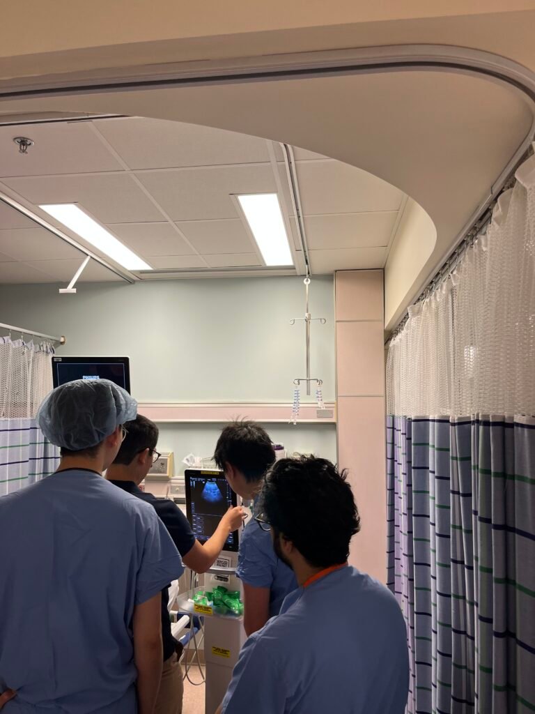
Attendees first examined a volunteer with an empty stomach to establish the baseline appearance of the gastric antrum. Faculty guided learners through probe positioning, optimization of image acquisition, and identification of key anatomical landmarks, including the left lobe of the liver, gastric antrum, pancreas, aorta, and inferior vena cava. This module established the normal reference examination that served as the foundation for subsequent modules. Faculty also demonstrated common scanning pitfalls and techniques to optimize image quality, allowing participants to recognize how subtle changes in probe position could significantly alter image interpretation. Emphasis was placed on developing a systematic scanning approach that learners could consistently reproduce in clinical practice.
Module 2. Model With Full Stomach (Clears)
Using the same volunteer, participants first reviewed the empty stomach examination and then repeated the scan immediately after ingestion of clear liquids. Comparing the two examinations in real time allowed learners to appreciate the characteristic ultrasound appearance of clear gastric contents and understand how gastric findings change following liquid intake. Faculty emphasized interpretation of these findings within the context of perioperative aspiration risk assessment. Participants were encouraged to identify the changes in antral appearance and discuss how these observations correlate with normal gastric physiology and current fasting recommendations. The real-time comparison reinforced the importance of integrating ultrasound findings with clinical history rather than relying solely on reported fasting intervals.
Module 3. Model With Full Stomach (Solids)
Participants evaluated a volunteer following ingestion of a standardized meal to identify the ultrasound appearance of solid gastric contents. Faculty encouraged comparison with the empty stomach and clear liquid examinations, reinforcing recognition of distinct imaging characteristics associated with each gastric content state. Discussion focused on how these findings influence aspiration risk assessment and perioperative planning. Participants reviewed qualitative features that distinguish solid from liquid gastric contents and discussed how these findings may alter anesthetic management in patients with uncertain fasting status or conditions associated with delayed gastric emptying. This comparative approach strengthened learners’ confidence in differentiating clinically relevant gastric content patterns during point-of-care assessment.
Module 4. GLP-1 and Management of Aspiration
This discussion-based module focused on integrating gastric ultrasound findings into perioperative decision-making through review of departmental quality improvement data and current clinical guidance. Faculty presented findings from an ongoing quality improvement initiative examining perioperative aspiration events within the department, using local incident reviews to identify clinical settings associated with a higher risk of aspiration and to illustrate how institutional experience has informed current practice. These data provided participants with practical insight into how aspiration events are recognized, analyzed, and translated into quality improvement initiatives. Building on this foundation, participants reviewed the department’s updated recommendations for perioperative management of patients receiving GLP-1 receptor agonists, including the rationale behind recent changes in practice. Faculty emphasized how gastric ultrasound can complement clinical assessment when aspiration risk is uncertain and discussed its role alongside patient history, fasting status, and procedural urgency when planning anesthetic care. Participants also received a concise reference guide summarizing the department’s recommendations for future clinical use.
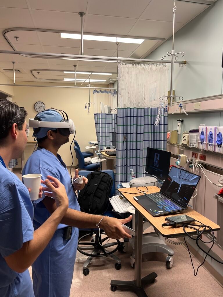
The workshop also included immersive virtual reality simulation developed by Vantari Systems. Attendees practiced probe orientation, spatial anatomy, and image acquisition in a three-dimensional environment that allowed real-time visualization of ultrasound images alongside probe movement. This simulation complemented the handson modules by providing additional opportunities to reinforce technical skills, improve anatomical understanding, and strengthen confidence in gastric ultrasound examination.
Module 5. Virtual Reality Gastric Ultrasound Simulation
The workshop also included immersive virtual reality simulation developed by Vantari Systems. Attendees practiced probe orientation, spatial anatomy, and image acquisition in a three-dimensional environment that allowed real-time visualization of ultrasound images alongside probe movement. This simulation complemented the handson modules by providing additional opportunities to reinforce technical skills, improve anatomical understanding, and strengthen confidence in gastric ultrasound examination.
Module 6. A Crisis
The workshop concluded with a high-fidelity simulation designed to integrate gastric ultrasound into perioperative crisis management. Set in the post-anesthesia care unit (PACU), the scenario required learners to evaluate aspiration risk as the patient’s condition evolved and to use gastric ultrasound findings, together with the clinical presentation, to guide perioperative decision-making. The simulation was conducted using a Rapid Cycle Deliberate Practice (RCDP) approach, allowing participants to receive immediate feedback, refine their management strategies, and reinforce key clinical actions through repeated practice. This module provided participants with an opportunity to apply the knowledge and skills developed throughout the workshop in a realistic, time-sensitive clinical setting.
CONCLUSION
The educational workshop provided attendees with a structured and interactive approach to learning perioperative gastric ultrasound through progressive, clinically relevant experiences. By combining image acquisition, interpretation of normal and pathologic findings, simulation, and evidence-based clinical discussion, learners strengthened both technical skills and clinical reasoning while building confidence in applying gastric ultrasound to perioperative decision-making. The integration of immersive technologies, departmental quality improvement initiatives, and simulation-based education further emphasized the practical application of gastric ultrasound in modern anesthetic practice. Educational programs that incorporate multiple complementary teaching strategies may enhance learner engagement while facilitating the translation of knowledge into perioperative patient care.