Atrial Fibrillation and R on T Phenomenon
Siraj Ahmad, MBBS
Feroze Mahmood, MD
Issue 3 | Volume 2 | August 2026


KEY TAKEAWAYS
- In atrial fibrillation the ventricular arrhythmia risk comes from the irregularity, not the rate, short–long– short sequences generate the R-on-T substrate beat to beat.
- The one cause of R-on-T fully under the clinician’s control is the shock itself – confirm synchronization markers on every complex before discharging, and correct potassium and magnesium.
- Timing is protective only while an organized rhythm persists. Once R-on-T has degenerated into ventricular fibrillation, unsynchronized defibrillation is the only option.
Introduction
The R-on-T phenomenon is a form of ventricular ectopy in which a premature ventricular depolarization occurs during the vulnerable phase of ventricular repolarization. Since this period represents a transition between refractory and excitable myocardium, an R-on-T beat can precipitate malignant ventricular arrhythmias.
Mechanism
During the T wave, ventricular recovery is heterogeneous, leaving some myocardial fibers refractory while others have regained excitability. If a premature ventricular impulse falls within this vulnerable window, it may encounter areas of conduction block and areas capable of conducting, promoting re-entry and potentially degenerating into polymorphic ventricular tachycardia or ventricular fibrillation.
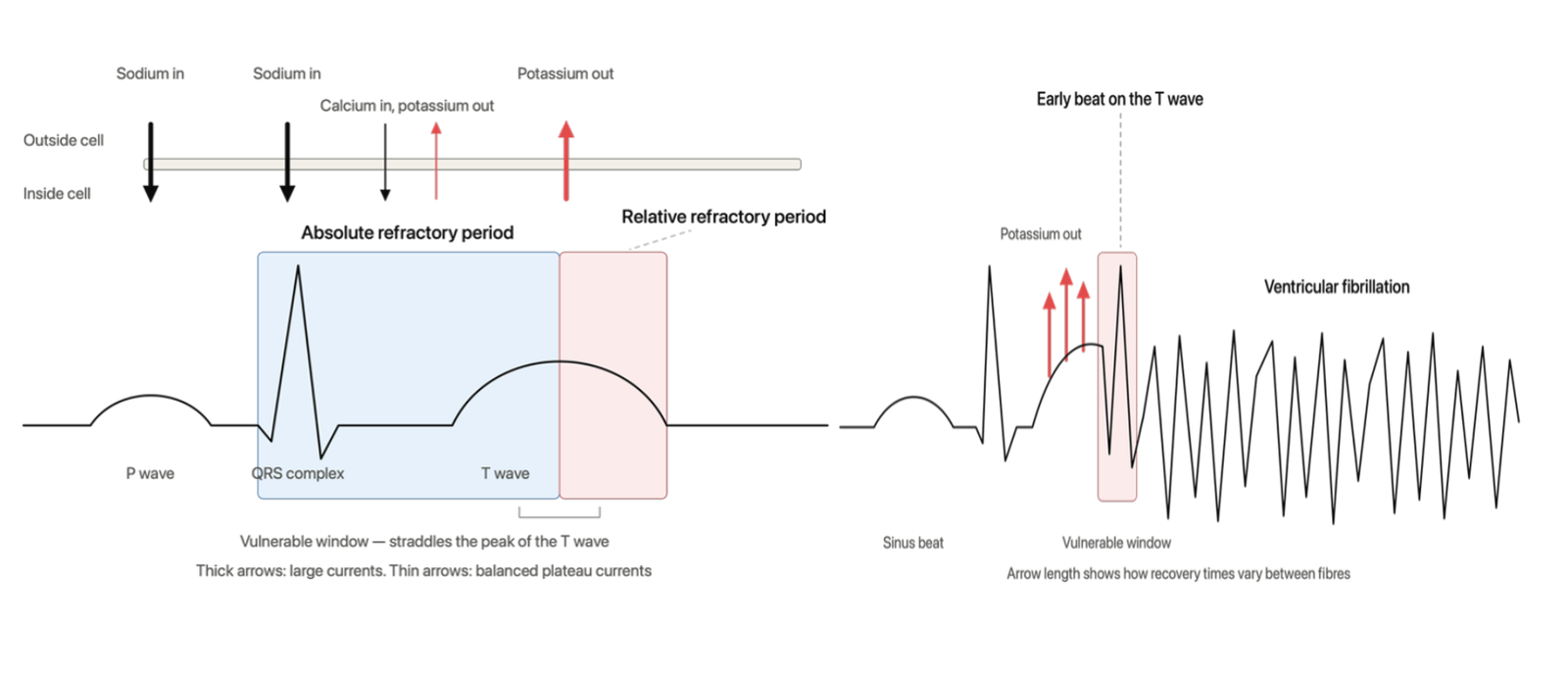
Left: Cardiac ion movements (Na⁺ influx, Ca²⁺ influx, K⁺ efflux) aligned with ECG refractory periods, highlighting the vulnerable period during repolarization (T wave).
Right: An early premature ventricular contraction (PVC) landing precisely on the vulnerable T-wave phase initiates disorganized ventricular fibrillation due to inhomogeneous tissue recovery.
Causes
Any depolarization arriving in the vulnerable window can cause it: a short-coupled ectopic, a paced beat following undersensing, or an ectopic in a heart with a prolonged QT interval from hypokalemia or hypomagnesemia. The important iatrogenic cause is a cardioversion shock delivered without synchronization, which lands wherever it falls in the cycle – including on the T wave.
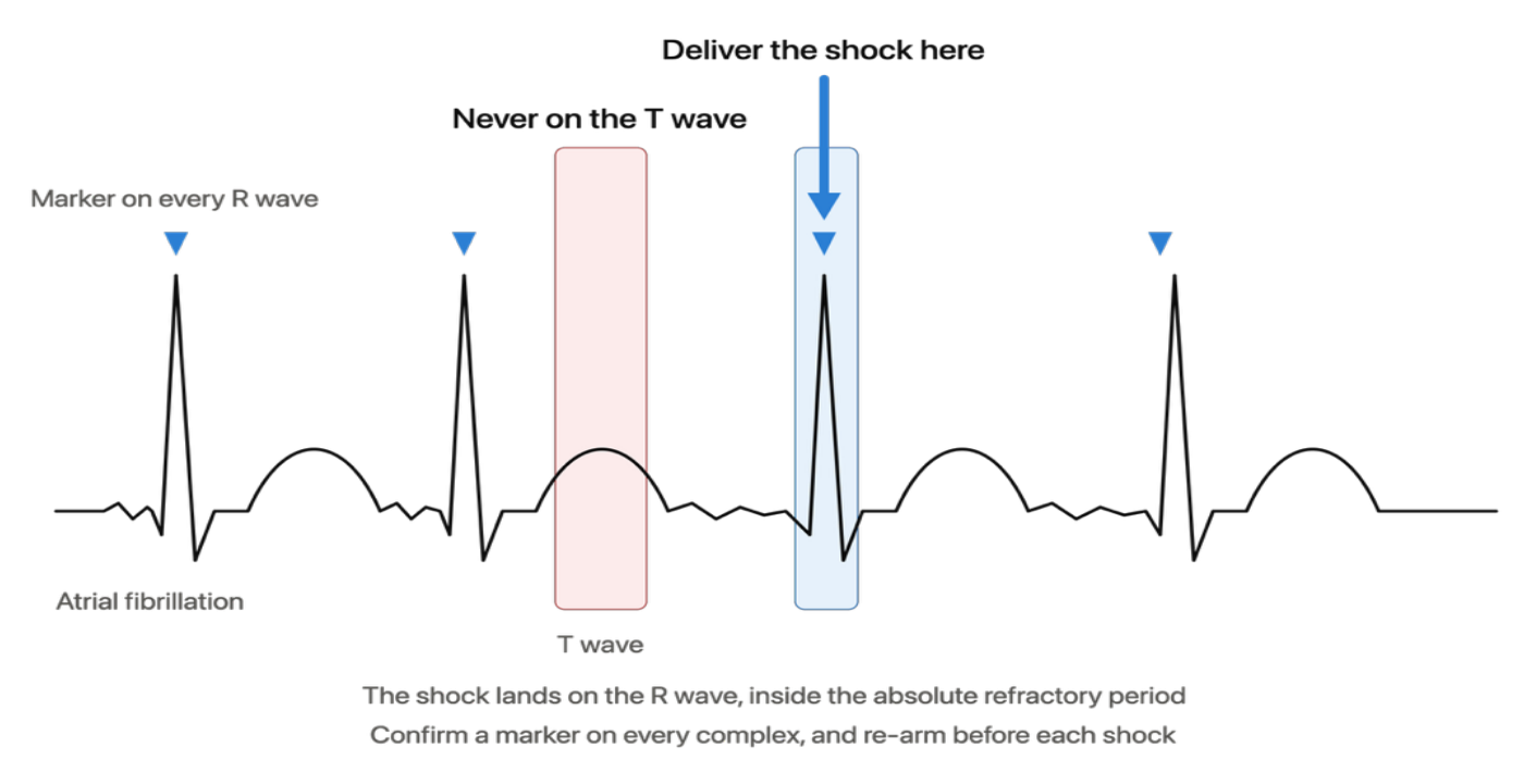
Illustration of an ECG tracing showing synchronized shock delivery on the R wave peak. Precise timing prevents electrical discharge during ventricular repolarization (T wave), preventing the R-on-T phenomenon and subsequent malignant arrhythmias.
Management
Prevention is the priority: restoring sinus rhythm, by synchronized cardioversion or pharmacological conversion, removes the short–long–short sequences that generate the substrate, whereas rate control leaves them intact. When performing electrical cardioversion, activating synchronization mode is vital to prevent the R-on-T phenomenon (Figured 2). Potassium and magnesium should be corrected, since a deficit widens the vulnerable window. Once ventricular fibrillation has developed, unsynchronized defibrillation is the only option.
REFERENCES
- Oksuz F, Sensoy B, Sahan E, et al. The classical “R-on-T” phenomenon. Indian Heart J 2015; 67: 392– 394.
- Qu Z, Liu MB, Olcese R, et al. R-on-T and the initiation of reentry revisited: Integrating old and new concepts. Heart Rhythm 2022; 19: 1369–1383.