A Percutaneous Alternative to Surgical Excision of Atrial Tumors
Fatima Noor, MBBS
Ruma Bose, MD
Issue 3 | Volume 2 | August 2026


KEY TAKEAWAYS
- Cardiac masses include both tumors and non-tumor conditions: They may represent benign or malignant tumors, thrombi, vegetations, or other lesions. Multimodality imaging is important for accurate diagnosis and treatment planning.
- Surgery remains the standard treatment for atrial myxomas: However, percutaneous removal may provide a less invasive option for carefully selected patients, particularly those at high surgical risk.
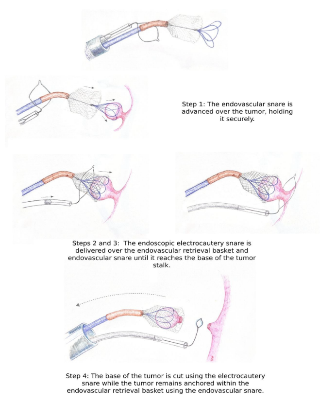
- The SEATTLE procedure combines tumor resection and retrieval: An electrocautery snare cuts the tumor stalk while an endovascular snare and basket secure the mass for removal through femoral venous access.
- The technique is promising but remains investigational: Early experience has shown that percutaneous removal is technically feasible, but larger studies are needed to establish its safety, long-term outcomes, and role compared with surgery
ABSTRACT
Cardiac masses include a broad range of neoplastic and non-neoplastic conditions, with myxoma representing the most common primary cardiac tumor. Surgical excision has long been standard of care. This review introduces the Simplified Extraction of Atrial Tumor with Targeted Loop Electricity (SEATTLE) procedure, a percutaneous technique that combines electrosurgical resection with endovascular retrieval to remove intracardiac masses without sternotomy. We provide a classification framework for cardiac masses and outline the technical steps of the SEATTLE procedure.
INTRODUCTION
Cardiac masses range from benign, incidentally discovered lesions to malignant tumors. Myxoma is the most common primary cardiac tumor in adults and typically arises from the interatrial septum. Surgical excision has been considered the definitive treatment. However, less invasive catheter-based techniques are emerging as potential alternatives. Understanding the broader classification of cardiac masses, along with minimally invasive treatment options, is increasingly relevant for trainees in cardiology, cardiac surgery, and structural heart disease. This review summarizes the classification of cardiac masses and introduces the SEATTLE procedure, a percutaneous technique for intracardiac tumor removal.
CLASSIFICATION OF CARDIAC MASSES
Cardiac masses are broadly divided into neoplastic and non-neoplastic categories (Table 1). Neoplastic masses are further subdivided into primary tumors — which may be benign or malignant — and metastatic tumors. Non-neoplastic masses, including thrombus and vegetations, are frequently encountered in clinical practice and must be distinguished from true tumors using multimodality imaging.
| Category | Examples | Typical location | Standard management | Gold standard imaging |
|---|---|---|---|---|
| Benign neoplasm | Myxoma, fibroma, lipoma, papillary fibroelastoma, rhabdomyoma, angioma, schwannoma, cystic tumor of the atrioventricular node | Left or right atrium (myxoma); ventricle and ventricular septum (fibroma); valve leaflets (fibroelastoma) | Surgical excision (traditional); percutaneous excision emerging for select myxomas | Echocardiography & cardiac MRI (myxoma); echocardiography (fibroelastomas); cardiac MRI (lipoma) |
| Malignant neoplasm | Angiosarcoma, rhabdomyosarcoma, myosarcoma, fibrosarcoma, malignant peripheral nerve sheath tumors | Right atrium (angiosarcoma); ventricles (rhabdomyosarcoma) | Surgery, chemotherapy, and/or palliation depending on stage | Cardiac MRI, biopsy |
| Metastatic tumor | Melanoma, lung, breast, renal cell carcinoma, lymphoma, leukemia | Epicardium, myocardium, endocardium | Treatment of primary malignancy; palliative measures | Cardiac MRI, cardiac CT, PET, biopsy |
| Thrombus | Left atrial or left atrial appendage thrombus, left ventricular thrombus | Left atrial appendage, left ventricle apex | Anticoagulation ± percutaneous or surgical removal | Cardiac MRI |
| Vegetation | Infective endocarditis | Valve leaflets | Antibiotics ± surgery | Transesophageal echocardiography |
| Pericardial cyst | Simple pericardial cyst | Cardiophrenic angle | Observation | Cardiac MRI, cardiac CT |
| Lipomatous hypertrophy | Lipomatous hypertrophy of the interatrial septum | Interatrial septum | Observation | Cardiac MRI |
TECHNIQUE AND PROCEDURAL STEPS
The Simplified Extraction Of Atrial Tumor with Targeted Loop Electricity, or SEATTLE, procedure was first described by Zachary L. Steinberg, James M. McCabe and colleagues in 2023 as a percutaneous approach to atrial myxoma removal. The technique pairs transcatheter electrosurgery with a dedicated endovascular retrieval system, avoiding the need for surgery. The procedure is performed under general anesthesia with continuous transesophageal echocardiographic guidance Vascular access is obtained, typically through the femoral vein. Echocardiographic imaging is used throughout the procedure to visualize the mass, confirm catheter position, and monitor for complications such as embolization or pericardial effusion. Once access to the tumor has been established, a steerable sheath is passed through the loop of an endoscopic electrocautery snare. An expandable endovascular retrieval system is then introduced through the steerable sheath, and an endovascular snare catheter is directed through the central lumen of the retrieval basket. The assembled system is pushed forward through the femoral venous sheath, with the electrocautery snare remaining outside and encircling the steerable sheath. Under echocardiographic guidance, the endovascular snare is passed over the atrial mass to capture and stabilize it. The expandable retrieval basket is then advanced over the mass to provide additional containment. Once the mass is secured, the electrocautery snare is advanced over the retrieval basket and endovascular snare to the base of the tumor stalk. The stalk is severed by retracting the electrocautery loop into its protective casing while delivering electrocautery energy, thereby separating the tumor from the atrial wall while it remains secured within the retrieval system. The detached tumor and retrieval basket are subsequently withdrawn together into the steerable sheath and removed through the femoral venous access. The extracted mass is then sent for pathological evaluation.
CONCLUSION
The SEATTLE procedure represents a percutaneous approach for the removal of selected atrial myxomas. This technique may offer patients an option that does not require sternotomy or cardiopulmonary bypass. Although the initial case demonstrated technical feasibility, further studies are needed to establish its safety, long-term outcomes, and role compared with conventional surgical excision.
REFERENCES
- Steinberg ZL, Elison D, Vincent LL, Oxorn D, McCabe JM. The Simplified Extraction of Atrial Tumor With Targeted Loop Electricity (SEATTLE) Procedure. JACC Case Rep. 2023;10:101758. Published 2023 Mar 15
- Pino PG, Moreo A, Lestuzzi C. Differential diagnosis of cardiac tumors: General consideration and echocardiographic approach. J Clin Ultrasound. 2022;50(8):1177- 1193.
- Reynen K. Cardiac myxomas. N Engl J Med. 1995;333(24):1610-1617.
- Angeli F, Bodega F, Bergamaschi L, et al. Multimodality imaging in the diagnostic work-up of patients with cardiac masses: JACC: CardioOncology State-of-the-Art Review. JACC CardioOncol. 2024;6(6):847-862. Published 2024 Oct 29