Optimization of Supraclavicular Block for Upper Extremity Anesthesia: Evidence-Based Approaches
Andrey Rakalin, MD
Published September 5, 2024 | Clinics in Medical Education
Issue 2 | Volume 1 | August 2024
A 35-year-old male presents with a distal radius fracture, as confirmed by X-ray. His vital signs are as follows: blood pressure 120/80 mmHg, heart rate 80 beats per minute, respiratory rate 16 breaths per minute, and oxygen saturation 98% on room air. Given these stable vital signs and the need for effective analgesia, which regional block would be most appropriate for this case?

Introduction
The supraclavicular block is useful for upper extremity surgery on the arm, elbow, forearm, wrist, and hand. The higher success rate and quicker onset of block has led to its name as “the spinal of the arm”. The bony and prominent clavicle and the curved contour can make imaging a challenge at the level of the base of the neck.
Anatomy
- The subclavian artery crosses over the first rib between the insertions of the anterior and middle scalene muscles, at mid-point of the clavicle.
- The brachial plexus can be seen as a bundle of hypoechoic round nodules just lateral and posterior to the artery.
- By changing the angle of the probe, the brachial plexus can appear as oval or flattened structure. It is usually 1-2cm deep. The first rib has echogenic shadow. Medial to the first rib there is echogenic pleura.
- The neurovascular bundle passes underneath the middle third of the clavicle.
- The brachial plexus at this level is usually located postero-lateral to the subclavian artery.
- The three trunks of the brachial plexus carry the entire sensory, motor and sympathetic innervation of the upper extremity, with exception of the uppermost part of the medial side of the arm (T2).
- This block is performed in the distal trunks and proximal divisions where the brachial plexus is most compact.It results in anesthesia of C5 to T1 dermatome. Both rib and pleura appear as hyperechoic linear area, but they can be differentiated as follows:
- There is a dark anechoic area under the first rib (acoustic shadow) and there is no movement with respiration. In contrast, the hyperechoic pleural surface is a brighter shimmering area that moves with respiration.
Position
- The patient can be placed in a semi-sitting or supine position with their head turned slightly away from the side in which the block will be placed.
- The arm is in neutral position with the shoulders relaxed.
- The patient can also be placed in the semi-fowlers position where the patient is lying supine but with the head of the bed raised at about 30o.
- Again, the head should be rotated away from puncture site and arm resting in a neutral position on the side with wrist supinated.
Preparation
Clean the injection site with chlorhexidine or iodine
Equipment
Typically, a 22G 50mm insulated needle is the most appropriate. Use a high frequency (>10MHz) linear probe to image the site.
Transducer
Place the probe in a transverse orientation on the neck, just superior to the clavicle at midpoint. It should point caudally.
Technique
For scanning above the clavicle, a linear probe is placed parallel to the clavicle and the needle is introduced laterally towards the midline, in-plane. The brachial plexus can be scanned and followed across the cervical region from its supraclavicular position and then cephalad to the interscalene groove. The transducer is tilted slightly inferiorly rather than perpendicular to the shin. Your Position: Next to the patient, close to the shoulder. To perform the block, advance the needle from lateral aspect via in-plane approach. After negative aspiration inject 20-25cc of local injection. Avoid high resistance to injection to reduce the risk of intraneural injection. The mean anesthetic volume used is 20-30 mL for single injection. The local anesthetic spreads around brachial plexus, lateral and superficial to the subclavian artery. This is adequate for anesthesia or the upper limb below the shoulder.

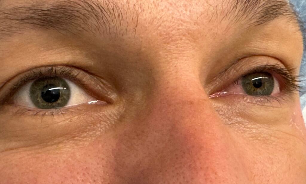
Figure 2
Horner Syndrome
Complications
- Phrenic nerve block
- Horner’s syndrome
- Recurrent laryngeal nerve paralysis
- Vascular puncture
- Pneumothorax
- Neurological deficits
Relative Contraindications
Relative contraindications include coagulopathy or respiratory compromise because of the risk of pneumothorax and phrenic nerve paralysis.
Absolute Contraindications
Absolute contraindications include patient refusal, allergy to local anesthetics, and infection or cellulitis at the injection site.
Clinical Pearls
When the needle is introduced under ultrasound guidance, the artery must be visible at all times. Identify the first rib and differentiate it from the pleural surface before needle advancement Needle tip should not disappear below the clavicle and should not be advanced medial to the artery. It is necessary to make small readjustments of needle tip position to ensure local anesthetic spread to the 3 trunks. Injection of local anesthetic will result in swelling of the sheath and separation of the nerves. The medial skin of the upper arm (intercostobrachial, T2) is not anesthetized and therefore it may need to be blocked separately. The musculocutaneous nerve is blocked to cover the medial skin of the upper limb.
Quiz Yourself
We have compiled cases for quick review of ECG and rhythm interpretations for efficient learning and skill enhancement.