Perioperative Challenges in Advanced Diastolic Dysfunction: A Cardiac Amyloidosis Case
Meghana Badari Narayan, MD
Issue 3 | Volume 2 | August 2026

KEY TAKEAWAYS
- Diastolic dysfunction is a common yet challenging comorbidity.
- Preoperative DD is an independent predictor of serious complications in non-cardiac surgery, increasing risk for postoperative pulmonary edema and major cardiac events in low and intermediate-risk surgeries.
- Based on the severity of DD, it is divided into grades which represent a physiological continuum of the disease.
- These patients have the highest perioperative haemodynamic risk. They may be preload dependent but simultaneously volume intolerant.
- Across all grades, perioperative management should minimize tachycardia, preserve sinus rhythm when possible, prevent myocardial ischaemia, avoid abrupt changes in afterload and use carefully titrated fluid therapy.
- Anesthetic management in restrictive cardiomyopathy requires careful understanding of physiology of diastole and pathophysiology of diastolic dysfunction.
CASE REPORT
A 44-year-old male presented with acute decompensated heart failure secondary to cardiac amyloidosis, with moderate global systolic dysfunction, grade III diastolic dysfunction, and a restrictive filling pattern. He required multiple vasopressors and continuous veno-venous hemofiltration for hemodynamic support. He underwent extracorporeal membrane oxygenation cannulation, complicated by lower extremity compartment syndrome requiring fasciotomy and venous cannula repositioning. He subsequently underwent cardiac transplantation. Diastolic dysfunction is a common yet challenging comorbidity. Anesthetic management in restrictive cardiomyopathy requires careful understanding of physiology of diastole and pathophysiology of diastolic dysfunction.
INTRODUCTION
Diastolic dysfunction (DD) represents a highly prevalent yet frequently underdiagnosed cardiac condition with significant clinical implications, serving as a major contributor to heart failure with preserved ejection fraction (HFpEF). LVDD is common in the general population, affecting approximately one-quarter of adults and 25%-30% of middle-aged and older adults in U.S. community studies. In contrast, HFpEF represents the symptomatic clinical manifestation of cardiac dysfunction and has an estimated general-population prevalence of approximately 1%-5% worldwide and 1%-1.5% in the United States. HFpEF affects up to 32 million people globally and approximately 3–3.5 million Americans, accounting for about half of all heart-failure cases.
The prognosis for HFpEF is poor despite a preserved LVEF, with a 21% 30-day all-cause hospital readmission rate and a 1-year mortality approaching 20% to 29%, comparable with patients with heart failure and reduced ejection fraction (HFrEF). Preoperative DD is an independent predictor of serious complications in non-cardiac surgery, increasing risk for postoperative pulmonary edema and major cardiac events in low and intermediate-risk surgeries. In high-risk vascular patients, DD is associated with significantly increased length of hospital stay and, even when asymptomatic, is a predictor of 30-day cardiovascular events and long-term mortality.
PHYSIOLOGY OF DIASTOLE
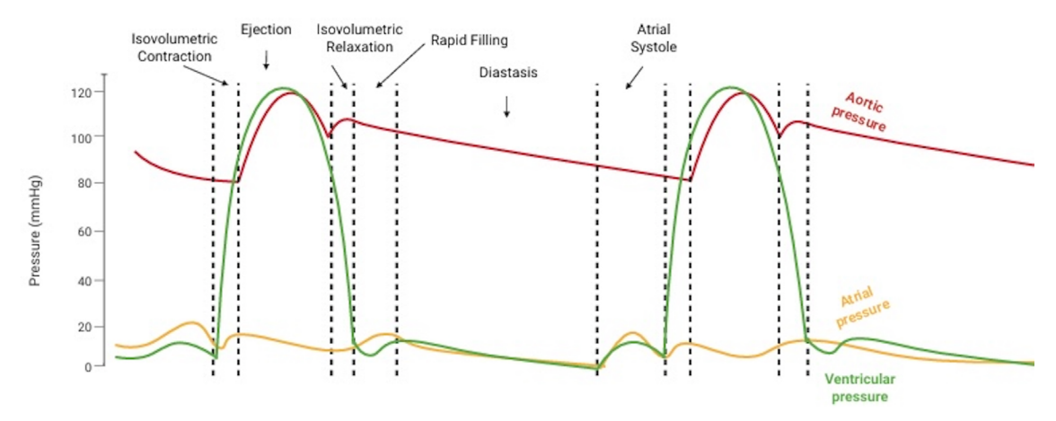
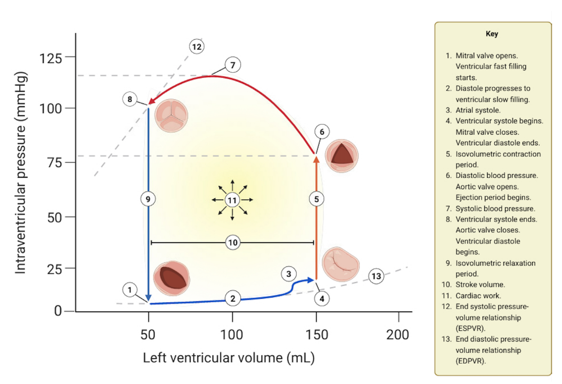
The diastole of the cardiac cycle encompasses the time between the closure of the aortic valve and the closure of the mitral valve. From a clinical perspective, normal diastolic function refers to the LV’s ability to relax and fill with blood, producing a sufficient stroke volume at low pressures. The ventricle’s ability to fill depends on its fixed viscoelastic stiffness and its variable capacity to relax.
Diastole consists of four distinct phases: isovolumetric relaxation, where LV pressure drops rapidly while volume remains constant; rapid filling, triggered when the mitral valve opens and blood rushes into the LV due to active ventricular suction; diastasis, a transitional pause of minimal passive filling as atrial and ventricular pressures equalize; and atrial contraction, where the left atrium actively contracts to complete ventricular filling before the mitral valve closes.
PATHOPHYSIOLOGY OF DIASTOLIC DYSFUNCTION
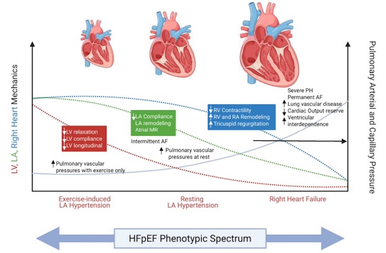
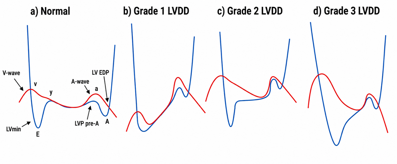
DD refers to impaired relaxation or filling of the ventricles during the diastolic phase of the cardiac cycle. Increased left ventricular filling pressure (LVFP) is the principal pathophysiological manifestation of LVDD. It arises from impaired myocardial relaxation, increased ventricular stiffness, or both, driven by intrinsic cardiomyocyte abnormalities, extracellular matrix remodeling, and maladaptive neurohumoral and endothelial signaling. Based on the severity of DD, it is divided into grades which represent a physiological continuum of the disease.
Grade 1: impaired relaxation with relatively preserved compliance.
The earliest abnormality is delayed or incomplete active myocardial relaxation. Ventricular recoil and early diastolic suction are reduced, so the ventricle fills more slowly during early diastole. Passive compliance is usually still relatively preserved, and mean left atrial pressure is generally normal at rest. Filling is redistributed toward late diastole, making the ventricle increasingly dependent on effective left atrial contraction to achieve an adequate end-diastolic volume.
At rest, this compensatory mechanism may preserve stroke volume and the patient may remain asymptomatic. During tachycardia, however, diastole shortens and the slowly relaxing ventricle may not have enough time to fill. Exercise, hypertension, myocardial ischaemia or increased circulating volume may consequently raise filling pressure despite normal resting pressure. The major perioperative considerations are tachycardia, loss of sinus rhythm and reduced venous return. Loss of atrial contraction, particularly with atrial fibrillation, can substantially reduce ventricular filling and stroke volume. Anaesthetic induced venodilation may decrease preload and produce hypotension despite preserved ejection fraction. When true hypovolaemia is present, judicious fluid replacement may improve filling; however, indiscriminate fluid loading can push the patient toward elevated filling pressures.
Grade 2: impaired relaxation with reduced compliance and elevated filling pressure
In grade 2 dysfunction, impaired relaxation persists, but passive ventricular compliance begins to deteriorate. The left atrium must generate a higher pressure to fill the increasingly stiff ventricle. This elevated atrial pressure restores the early atrioventricular pressure gradient and maintains ventricular filling despite poor ventricular suction. This is the physiological basis of the term pseudonormalization. The higher pressure is transmitted backward to the pulmonary veins. Chronic pressure loading promotes left atrial remodelling, impaired atrial reserve and contractile function, pulmonary venous hypertension and, with progression, post-capillary pulmonary hypertension. Stroke volume may remain normal at rest, but cardiac reserve is reduced because increasing filling volume produces elevated filling pressure rather than an appropriate increase in end-diastolic volume.
Patients have a particularly narrow volume tolerance. With fluid excess, left atrial pressure rises rapidly, producing pulmonary congestion. Tachycardia, atrial fibrillation, systemic hypertension and myocardial ischaemia further increase filling pressures. If hypotension after induction is treated with repeated fluid boluses it can correct the blood pressure while worsening pulmonary congestion. Fluid administration should be incremental and guided by repeated assessment rather than by blood pressure alone. For major surgery or anticipated large fluid shifts, closer haemodynamic monitoring is reasonable.
Grade 3: restrictive physiology with markedly elevated filling pressure
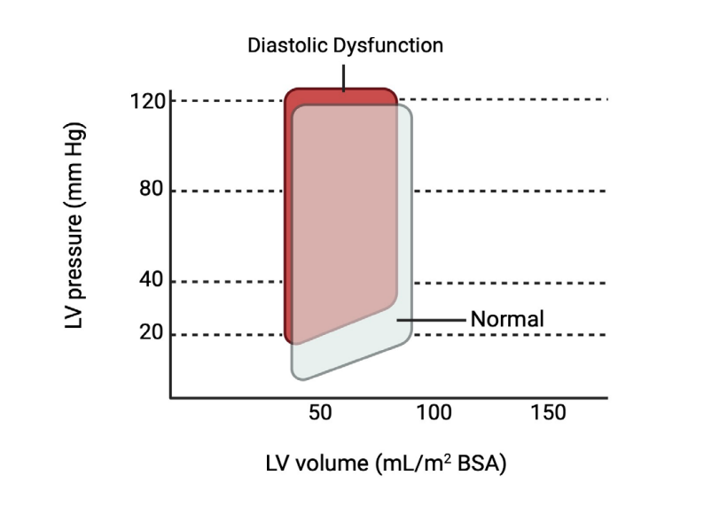
Grade 3 represents advanced diastolic dysfunction. Ventricular compliance is severely reduced, myocardial relaxation is markedly abnormal and left atrial pressure is substantially elevated. The stiff ventricle accepts most of its filling rapidly during early diastole because of the high atrioventricular pressure gradient, but filling terminates abruptly as ventricular pressure rises quickly and equals left atrial pressure. The end-diastolic pressure-volume relationship is markedly shifted upward and leftward. Consequently, a small increase in ventricular volume can produce a large increase in LV end-diastolic and left atrial pressures. Pulmonary venous hypertension, pulmonary oedema, post-capillary pulmonary hypertension and secondary right ventricular dysfunction may develop. Cardiac output becomes relatively fixed because the ventricle cannot meaningfully recruit the Frank-Starling mechanism without an excessive rise in filling pressure.
These patients have the highest perioperative haemodynamic risk. They may be preload dependent but simultaneously volume intolerant. Induction related vasodilation can cause profound hypotension even when left-sided filling pressures are already high. Fluid boluses can rapidly precipitate pulmonary edema. Tachycardia, hypertension, atrial arrhythmias, myocardial ischaemia and abrupt sympathetic stimulation can produce acute decompensation. Positive-pressure ventilation may reduce venous return, whereas emergence and spontaneous breathing increase venous return, afterload and sympathetic tone. Fluid shifts after surgery can produce delayed pulmonary edema even when the intraoperative course appears acceptable. Vasopressor and, when appropriate, inotropic support may be preferable to volume administration. An arterial line and perioperative echocardiographic or advanced haemodynamic assessment should be considered for high-risk procedures, substantial blood loss or major fluid shifts.
Across all grades, perioperative management should minimize tachycardia, preserve sinus rhythm when possible, prevent myocardial ischaemia, avoid abrupt changes in afterload and use carefully titrated fluid therapy. Adrenergic surges during laryngoscopy, surgical stimulation, pain, shivering and extubation can worsen relaxation and increase filling pressures. Postoperative monitoring is particularly important because sympathetic activation and redistribution of interstitial fluid into the circulation may unmask congestion.
REFERENCES
- Ottosen CI, Nadruz W, Inciardi RM, Johansen ND, Fudim M, Biering-Sørensen T. Diastolic dysfunction in hypertension: a comprehensive review of pathophysiology, diagnosis, and treatment. Eur Heart J Cardiovasc Imaging. 2024;25(11):1525-1536. doi:10.1093/ehjci/jeae178
- Fisher T, Abbawy M, Holden F, Cotton J, Hothi S, Ingram T, et al. Diastolic dysfunction in acute and critical illness: acute pathophysiology to chronic heart failure. JACC Adv. 2026;5(2):102532. doi:10.1016/j.jacadv.2025.102532.
- Vlasopoulou K, Synetos A, Ktenopoulos N, et al. Unmasking left ventricular diastolic dysfunction: pathophysiology, diagnosis, and treatment strategies. Med Sci (Basel). 2025;13(3):204. doi:10.3390/medsci13030204.
- Borlaug BA, Sharma K, Shah SJ, Ho JE, Santhanakrishnan R, Beussink-Nelson L, et al. Heart failure with preserved ejection fraction: JACC Scientific Statement. J Am Coll Cardiol. 2023 May 9;81(18):1810-1834. doi: 10.1016/j.jacc.2023.01.049.
- Cios TJ, Klick JC, Roberts SM. Managing diastolic dysfunction perioperatively. Semin Cardiothorac Vasc Anesth. 2023;27(1):38-48. doi:10.1177/10892532221142441.