Rethinking Blood Pressure Management During Carotid Endarterectomy: The Potential Role of Blood Pressure Variability
Yifan Bu, MD
Issue 3 | Volume 2 | August 2026

BACKGROUND
Perioperative stroke remains one of the most severe complications following carotid endarterectomy (CEA). However, optimal intraoperative blood pressure management remains incompletely defined. Current practice is largely guided by physiologic reasoning rather than high-level evidence. During carotid cross-clamping, cerebral perfusion of the ipsilateral hemisphere shifts from an autoregulated state to a collateral-dependent, pressure-passive circulation, where cerebral blood flow becomes highly dependent on systemic arterial pressure. In this setting, anesthesiologists play a critical role in maintaining adequate cerebral perfusion by manipulating blood pressure, ventilation, and vasoactive medications.
A PHASE-SPECIFIC HEMODYNAMIC STRATEGY
Current guidelines therefore advocate a phase-specific hemodynamic strategy rather than a single universal blood pressure target. Blood pressure augmentation during carotid clamping is widely adopted because increased systemic pressure improves collateral perfusion through the Circle of Willis, increases middle cerebral artery flow velocity, and may reverse potential clamp-induced cerebral ischemia. After unclamping, restoration of perfusion to chronically vasodilated cerebral vessels increases the risk of cerebral hyperperfusion, while thromboembolism arising from the endarterectomy site becomes the dominant mechanism of perioperative stroke. Consequently, blood pressure is generally returned toward baseline after reperfusion to minimize hyperperfusion-related injury.
UNCERTAINTY IN OPTIMAL BLOOD PRESSURE TARGETS
Despite widespread agreement that blood pressure is critical, the optimal target remains uncertain. Existing recommendations are supported primarily by observational studies and physiologic experiments, with no completed randomized trial establishing evidence-based intraoperative blood pressure targets for CEA. Increasing evidence also suggests that relative blood pressure change, rather than an absolute MAP threshold, better reflects cerebral perfusion in patients with chronic carotid stenosis, whose autoregulatory curves are frequently shifted and collateral reserve varies between individuals. Ongoing studies investigating autoregulation-guided blood pressure management further highlight the limitations of fixed MAP thresholds and the movement toward individualized hemodynamic management.
BLOOD PRESSURE VARIABILITY: AN UNDEREXPLORED PHENOTYPE
An equally important but considerably less explored question is whether blood pressure variability (BPV), rather than mean blood pressure alone, contributes to perioperative cerebral injury. BPV has emerged as an important cardiovascular risk marker in hypertension and perioperative medicine, but its role during CEA remains poorly understood. Current evidence is remarkably sparse: only one CEA-specific study has directly evaluated intraoperative BPV, using postoperative headache rather than stroke as the primary outcome, while larger registry studies (based on the VQI data set) describe postoperative “hemodynamic lability” without calculating formal variability indices. Moreover, nearly all available studies reduce an entire operation to a single BPV value, ignoring the fundamentally different physiology of the dissection, cross-clamp, and post-unclamp phases. Such whole-procedure metrics cannot distinguish appropriate protocol-driven blood pressure modulation from pathological instability. Important methodological issues, including lack of standardized BPV metrics, dependence on sampling frequency, inability to separate spontaneous instability from vasoactive intervention, and absence of phase-specific analyses, remain unresolved.
STUDY DESIGN
To address these limitations, we analyzed high-resolution intraoperative arterial pressure waveforms from 95 consecutive carotid endarterectomy patients, including more than 200,000 arterial pressure measurements, with blood pressure trajectories normalized into pre-clamp, clamp, and post-unclamp phases. Rather than focusing solely on mean arterial pressure, we characterized BPV using 5 indices, including SD, coefficient of variation, average real variability, RMSSD, and interquartile range, to capture multiple dimensions of hemodynamic instability.
RESULTS
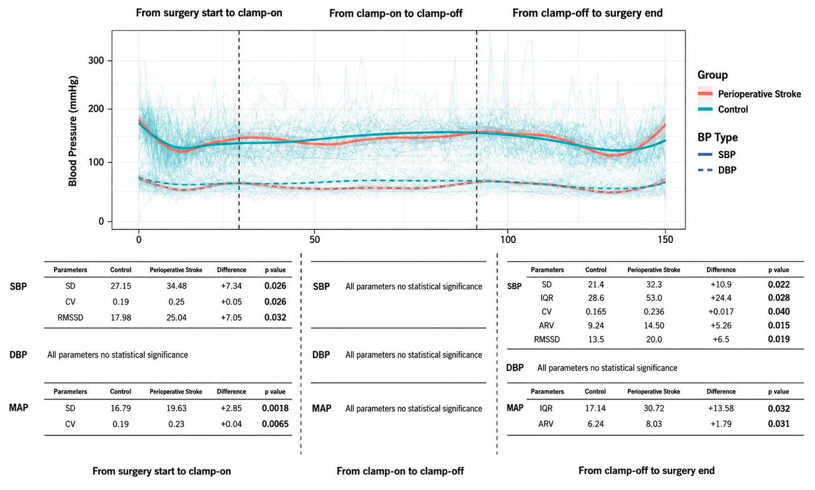
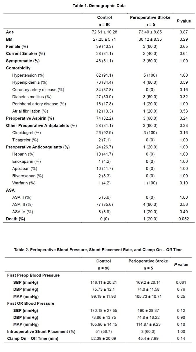
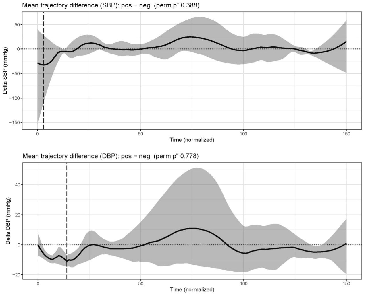
Although baseline characteristics, initial blood pressure, clamp duration, and shunt utilization were similar between patients with and without perioperative stroke (Fig. 1, see companion figures document), mixed-effects and trajectory analyses demonstrated different temporal blood pressure dynamics. Mean blood pressure trajectories alone were not consistently different throughout the procedure (Fig. 2, see companion figures document). In contrast, stroke patients exhibited a reproducible pattern of increased BPV, particularly involving systolic blood pressure and MAP during the pre-clamp and post-unclamp periods, whereas variability during carotid clamping remained relatively similar between groups (Fig. 3, see companion figures document). These findings were consistent across multiple complementary BPV metrics, suggesting that perioperative stroke may be associated with a distinct high-BPV hemodynamic phenotype, independent of differences in mean blood pressure.
MECHANISTIC CONSIDERATIONS
These observations raise several important mechanistic questions. Current concepts of cerebral protection during CEA largely emphasize maintaining adequate collateral perfusion during carotid clamping. However, contemporary hemodynamic studies suggest that cerebral circulation after clamping represents a dynamically reconfigured collateral network rather than simply a low-flow state. Blood pressure fluctuations may therefore influence not only cerebral perfusion but also collateral flow dynamics and embolic transport after reperfusion. The post-unclamp period is particularly intriguing because embolic events become the predominant cause of perioperative stroke while cerebral vessels remain vulnerable to hemodynamic instability. Whether increased BPV alters embolic washout, collateral flow efficiency, or regional cerebral perfusion remains unknown, but these mechanisms provide biologically plausible explanations linking hemodynamic instability with cerebral injury.
Our findings therefore should not be interpreted as establishing a causal relationship between BPV and perioperative stroke. Rather, they identify BPV as a previously underrecognized hemodynamic phenotype that may capture aspects of cerebral physiology not reflected by mean arterial pressure alone.
CLINICAL IMPLICATIONS
Although BPV cannot yet be regarded as a therapeutic target, the available evidence suggests several practical strategies that may improve hemodynamic stability during carotid endarterectomy.
First, continuous invasive arterial pressure monitoring should be maintained throughout all operative phases. Unlike intermittent cuff measurements, continuous arterial waveforms permit recognition of short time scale fluctuations that characterize BPV and provide the foundation for timely intervention.
Second, continuous vasopressor infusion may be preferable to repeated manual boluses in patients at high risk of hemodynamic instability. Randomized studies in noncardiac surgery have demonstrated that continuous norepinephrine infusion reduces short-term MAP variability, quantified using generalized average real variability, while preserving cardiac output and microcirculatory perfusion more effectively than intermittent bolus administration.
Third, blood pressure management during CEA should remain phase-specific rather than threshold-based. Current evidence supports maintaining augmented perfusion pressure during carotid clamping while avoiding excessive hypertension after unclamping, when embolic injury and cerebral hyperperfusion become the dominant concerns. Rather than targeting a single MAP value, anesthesiologists should consider maintaining blood pressure within an individualized target band appropriate for each operative phase.
Current evidence does not clearly favor one vasopressor over another for routine management during CEA. The only randomized CEA-specific comparison demonstrated similar cerebral oxygenation after phenylephrine and ephedrine, whereas newer physiological studies in collateral-dependent cerebrovascular disease suggest that norepinephrine may better preserve cardiac output and cerebral oxygen delivery. These observations remain hypothesis-generating, and vasopressor selection should currently be individualized according to the patient’s cardiovascular physiology rather than presumed cerebrovascular superiority of any single agent.
FUTURE DIRECTIONS
Future investigations should move beyond conventional mean blood pressure targets toward phase-specific hemodynamic management that integrates blood pressure trajectories, collateral physiology, and individualized cerebral autoregulation. Within this framework, BPV should be viewed not merely as a statistical descriptor of instability, but as a potentially modifiable physiological phenotype reflecting the interaction between anesthetic management and collateral cerebral circulation.
REFERENCES
- AbuRahma, A.F., Avgerinos, E.D., Chang, R.W., et al., 2022. The Society for Vascular Surgery implementation document for management of extracranial cerebrovascular disease. Journal of Vascular Surgery, 75(1S), pp.26S–98S.
- Benesch, C., Glance, L.G., Derdeyn, C.P., et al., 2021. Perioperative neurological evaluation and management to lower the risk of acute stroke in patients undergoing noncardiac, nonneurological surgery: a scientific statement from the American Heart Association/American Stroke Association. Circulation, 143(19), pp.e923–e946.
- Heyer, E.J., Mergeche, J.L., Anastasian, Z.H., et al., 2014. Arterial blood pressure management during carotid endarterectomy and early cognitive dysfunction. Neurosurgery, 74(3), pp.245–251.
- Zipfel, J., Bantle, S.J., Magunia, H., et al., 2020. Non-invasive cerebral autoregulation monitoring during awake carotid endarterectomy identifies clinically significant brain ischaemia. European Journal of Vascular and Endovascular Surgery, 60(5), pp.647–654.
- Spencer, M.P., Thomas, G.I. and Moehring, M.A., 1992. Relation between middle cerebral artery blood flow velocity and stump pressure during carotid endarterectomy. Stroke, 23(10), pp.1439–1445.
- Wang, J., Guo, L., Holdefer, R.N., et al., 2021. Intraoperative neurophysiology and transcranial Doppler for detection of cerebral ischemia and hyperperfusion during carotid endarterectomy. World Neurosurgery, 154, pp.e245–e253.
- Rots, M.L., Fassaert, L.M.M., Kappelle, L.J., et al., 2020. Intra-operative hypotension is a risk factor for post-operative silent brain ischaemia in patients with pre-operative hypertension undergoing carotid endarterectomy. European Journal of Vascular and Endovascular Surgery, 59(4), pp.526–534.
- Lee, D., Batista, P.M., McMackin, K.K., et al., 2022. Intraoperative blood pressure lability carries a higher risk of headache after carotid endarterectomy. Journal of Vascular Surgery, 75(2), pp.592–598.e1.
- Kobayashi, M., Ogasawara, K., Yoshida, K., et al., 2011. Intentional hypertension during dissection of carotid arteries in endarterectomy prevents postoperative development of new cerebral ischemic lesions caused by intraoperative microemboli. Neurosurgery, 69(2), pp.301–307.
- Wang, G.J., Beck, A.W., DeMartino, R.R., et al., 2017. Insight into the cerebral hyperperfusion syndrome following carotid endarterectomy from the National Vascular Quality Initiative. Journal of Vascular Surgery, 65(2), pp.381–389.e2.